
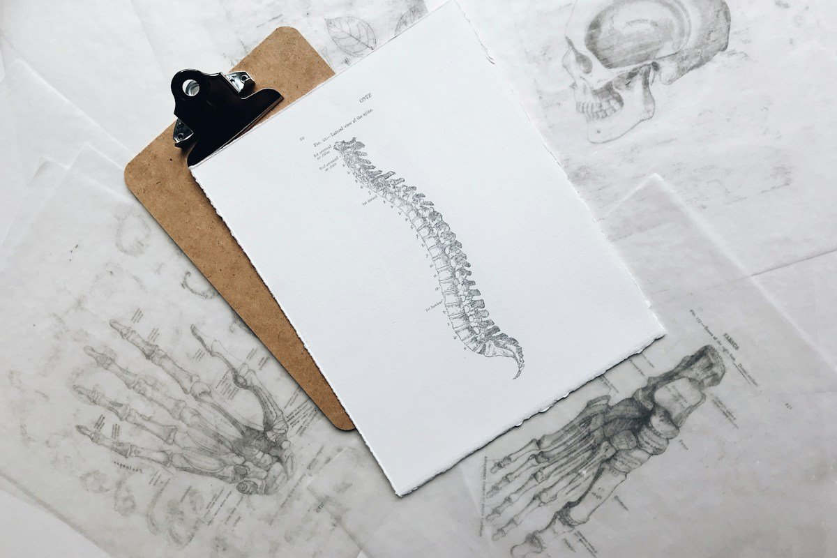

Sedentary occupational ergonomics represents one of the most pervasive yet underestimated biomechanical disruptions confronting modern industrialized workforces. Prolonged unbroken seated postures, characterized by sustained hip flexion, thoracic kyphosis, cervical protraction, and scapular downward rotation, systematically distort evolutionary human musculoskeletal design. Rather than functioning as dynamic, multi-directional bipedal organisms optimized for locomotion and load carriage, desk-bound knowledge workers subject their passive connective tissues and active myofascial structures to hours of static, low-load muscular ischemia.
Over months and years, this sustained postural stasis alters length-tension relationships across reciprocal muscle groups, drives persistent ligamentous creep, diminishes joint lubrication, and compromises neurological motor recruitment patterns. The resulting musculoskeletal dysfunctions do not merely present as transient localized discomfort; they manifest as chronic spinal disc compression, chronic tension headaches, temporomandibular joint pain, lumbo-pelvic instability, and accelerated structural degeneration of the axial skeleton.
Reversing these chronic postural distortions requires far more than passive ergonomics adjustments, ergonomic office chairs, or generic stretching routines. Effective musculoskeletal restoration necessitates a systematic clinical biomechanics framework: identifying specific neuro-muscular inhibition patterns, restoring physiological joint range of motion, realigning the lumbo-pelvic cylinder, retraining the inner core stabilizing musculature, and integrating active corrective exercise sequences into daily professional routines.
This comprehensive biomechanical guide delivers an authoritative, evidence-based roadmap for postural restoration tailored specifically to sedentary professionals, physical therapists, and movement specialists. By bridging functional anatomical science, joint kinematics, and corrective kinesiology, this clinical manual provides the diagnostic methodologies, progressive movement protocols, and ergonomic strategies necessary to reverse postural dysfunction, eliminate chronic musculoskeletal pain, and re-establish resilient physiological posture.
Functional Biomechanics of Prolonged Seated Postures: Kinematic Cascades
Human posture is maintained through continuous, subconscious proprioceptive feedback loops involving mechanoreceptors in joint capsules, Golgi tendon organs, muscle spindles, and the vestibular system. When an individual adopts a seated position for prolonged periods, the pelvis shifts into a posterior pelvic tilt, flattening the natural lumbar lordotic curvature. This flattening shifts axial gravitational loading away from the posterior facet joints and distributes massive hydrostatic shear stresses directly onto the anterior lumbar intervertebral discs.
Simultaneously, the sustained ninety-degree flexion angle of the hips places the primary hip flexor musculature, specifically the psoas major, iliacus, and rectus femoris, in a chronically shortened position. Through the physiological principle of adaptive shortening, muscle sarcomeres in series are lost, increasing passive muscle stiffness. Via reciprocal inhibition, hypertonic hip flexors transmit continuous inhibitory neurological signals to their direct functional antagonists: the gluteus maximus and gluteus medius.
This functional neurological inhibition, clinically termed gluteal amnesia, robs the lumbo-pelvic-hip complex of its primary posterior kinetic driver. When the gluteal complex fails to activate effectively during standing or walking, synergistic muscle groups—such as the hamstrings, adductor magnus, and lumbar erector spinae—are forced to compensate for hip extension, leading to severe synergistic dominance, chronic hamstring strain, and lumbo-sacral shearing.
Ascending the kinetic chain, the loss of lumbar lordosis triggers a reciprocal thoracic compensation. The thoracic spine collapses into exaggerated kyphosis, which drives the scapulae into lateral abduction, anterior tilting, and downward rotation. This structural misalignment shortens the pectoralis minor and subclavius muscles, while over-lengthening and neurologically inhibiting the middle trapezius, lower trapezius, and rhomboids, establishing the anatomical foundation for chronic upper quarter pain.
Upper Crossed Syndrome: Neuro-Myofascial Imbalance in the Cervical Spine
First codified by Czech physiatrist Dr. Vladimir Janda, Upper Crossed Syndrome (UCS) describes a predictable pattern of neuro-myofascial imbalance across the dorsal and ventral aspects of the shoulder girdle and cervical spine. In sedentary computer workers, this cross-pattern is driven by hours of visual focus on monitors paired with typing or mousing postures.
UCS is characterized by hypertonicity and shortening of the upper trapezius and levator scapulae on the dorsal side, crossing diagonally with tightness in the pectoralis major and pectoralis minor on the ventral side. Reciprocally, the deep cervical flexors (longus colli, longus capitis, and rectus capitis anterior) on the ventral side become neurologically inhibited and weak, crossing diagonally with weakness in the lower trapezius, middle trapezius, and serratus anterior on the dorsal side.
The most glaring clinical presentation of UCS is forward head posture (cervical protraction). For every single inch the head shifts anteriorly past the plumb line of the acromion process, the effective gravitational load carried by the cervical spine increases by approximately ten pounds. A computer operator displaying three inches of forward head posture subjects their posterior suboccipital and upper cervical musculature to forty to fifty pounds of continuous static mechanical strain.
This perpetual muscular traction compresses the greater and lesser occipital nerves, causing cervicogenic tension headaches radiating into the temporal and frontal regions. Furthermore, the anteriorly tilted scapula reduces the subacromial space, causing mechanical impingement of the supraspinatus tendon and subacromial bursa against the anteroinferior acromion during shoulder abduction, leading to chronic rotator cuff tendinopathy.
Lower Crossed Syndrome and Pelvic Tilt Kinematics: Lumbo-Pelvic Instability
Vladimir Janda equally defined Lower Crossed Syndrome (LCS) to delineate reciprocal muscular imbalances governing the lumbo-pelvic-hip complex. In desk workers, LCS manifests in two primary presentations: Type A (postural pelvic dysfunction involving dominant hip flexors and lumbar erectors) and Type B (abdominal and gluteal inhibition with posterior pelvic rotation).
In classic Type A presentation, hypertonic psoas, iliacus, rectus femoris, and tensor fasciae latae cross diagonally with overactive, shortened thoracolumbar erector spinae and quadratus lumborum. Crossing reciprocally are inhibited, hypotonic abdominal wall musculature (transversus abdominis, rectus abdominis, and internal obliques) and inhibited, weakened gluteus maximus and medius muscles.
This specific muscular imbalance forces the pelvis into an excessive anterior tilt during standing postures. Anterior pelvic tilt increases lumbar lordosis, jamming the posterior zygapophysial facet joints together and narrowing the neural foramina through which lumbar nerve roots exit. Over time, facet joint irritation triggers defensive muscular spasm in the lumbar paraspinals, creating a self-perpetuating pain-spasm-pain cycle.
In Type B presentation, commonly observed in slouched sitting postures, the pelvis rotates into excessive posterior tilt with complete flexion of the lumbar spine. This chronic flexion stretches the posterior longitudinal ligament, the interspinous ligaments, and the posterior annulus fibrosus beyond their physiological limits, inviting posterior and posterolateral lumbar disc herniations under minimal external loading.
Thoracic Spine Hypomobility and Rib Cage Mechanics
The thoracic spine is anatomically designed to provide rotational mobility and structural stability for thoracic cage organs. However, prolonged sitting in slouching positions locks the thoracic facets into continuous flexion, systematically extinguishing thoracic extension and rotation.
When the thoracic spine becomes rigid and hypomobile, the human kinetic chain must compensate elsewhere to achieve functional movements. In rotational tasks (such as turning to reach or glancing behind), the mobile cervical spine and the naturally rigid lumbar spine are forced to produce compensatory rotation. Because lumbar facet joints are aligned in the sagittal plane specifically to prevent axial rotation, compensatory lumbar twisting creates high shearing forces across lumbar discs, accelerating disc degradation.
Furthermore, thoracic kyphosis directly impacts respiratory mechanics. A collapsed thoracic cage compresses the abdominal viscera, preventing the diaphragm from descending fully during inspiration. Sedentary workers develop shallow, apical breathing patterns, relying on accessory respiratory muscles: the sternocleidomastoid, scalenes, and pectoralis minor. Overactive scalenes compress the brachial plexus and subclavian vessels against the first rib, producing neurovascular thoracic outlet syndrome (TOS) characterized by arm numbness, tingling, and grip weakness.
Scapular Dyskinesis: Glenohumeral Mechanics and Rotator Cuff Dysfunction
The scapula serves as the mobile mechanical platform for all glenohumeral movement. Normal arm elevation requires coordinated scapulohumeral rhythm: for every two degrees of glenohumeral elevation, the scapula must upwardly rotate by one degree, accompanied by posterior tilting and external rotation.
Sedentary desk posture disrupts this kinematic coordination, resulting in scapular dyskinesis. Tightness in the pectoralis minor pulls the coracoid process anteroinferiorly, causing persistent anterior tilting and internal rotation of the scapula. Concurrently, neurological inhibition of the serratus anterior and lower trapezius leaves the scapula unable to upwardly rotate smoothly along the thoracic wall.
When a sedentary worker reaches overhead or extends an arm to type, the acromion fails to clear the humeral head. The subacromial space narrows from an optimal ten millimeters to less than five millimeters, pinching the supraspinatus tendon and long head of the biceps brachii. This mechanical friction produces micro-tears, chronic inflammation, and eventual full-thickness rotator cuff rupture, even in the absence of acute sports trauma.
The Lumbo-Pelvic Cylinder: Diaphragm, Transversus Abdominis, and Pelvic Floor
Spinal stability is not generated by superficial muscles like the rectus abdominis or superficial erector spinae; it is maintained by the deep core stability mechanism: the lumbo-pelvic cylinder. This anatomical cylinder is bounded superiorly by the respiratory diaphragm, inferiorly by the pelvic floor musculature, circumferentially by the transversus abdominis, and posteriorly by the lumbar multifidus.
When functional, these four muscle groups contract co-operatively prior to any limb movement, regulating intra-abdominal pressure (IAP) to create a hydraulic stabilizing column that supports the lumbar spine from within. However, slumped sitting disrupts this synergistic co-activation: the flattened diaphragm cannot generate downward pressure, the abdominal wall is mechanically stretched and flaccid, and the pelvic floor experiences uneven chronic compression.
Deprived of internal intra-abdominal pressure stabilization, the spine relies entirely on passive bony and ligamentous structures to resist gravitational shear. When a sedentary individual suddenly bends forward to lift an everyday object, the deep stabilizing muscles fail to fire on time, allowing micro-instability and acute intervertebral disc injury.
Cervicogenic Cephalea and Craniofacial Biomechanics: TMJ Articulation
Forward head posture exerts a profound, destabilizing impact on the craniofacial complex and temporomandibular joint (TMJ) kinematics. As the cranium glides forward relative to the cervical spine, the mandible is pulled posteriorly and inferiorly by the passive tension of the suprahyoid and infrahyoid musculature. This mechanical traction retro-positions the condylar process of the mandible within the mandibular fossa of the temporal bone.
To compensate and prevent the mouth from falling open, the masseter and temporalis muscles undergo continuous hypertonic co-contraction. Overactive pterygoid musculature pulls the articular fibrocartilaginous disc of the TMJ anteriorly and medially. Over time, this anterior disc displacement creates chronic joint clicking, crepitus, severe masticatory pain, and nocturnal bruxism (teeth grinding) in high-stress desk workers.
Concurrently, chronic suboccipital shortening compresses the suboccipital nerve (C1 dorsal ramus) and the greater occipital nerve (C2 dorsal ramus) as they pierce the rectus capitis posterior major and obliquus capitis inferior muscles. This neurovascular impingement triggers cervicogenic headaches: throbbing, non-pulsatile pain that originates at the cranial base and radiates in a characteristic ram’s horn distribution over the vertex of the skull into the retro-orbital eye socket.
Neuro-Vascular Entrapment Syndromes: Thoracic Outlet and Sciatic Mobility
Static occupational postures create critical anatomical bottlenecks where major peripheral nerves and vascular bundles are vulnerable to mechanical entrapment and chronic ischemia. In the upper quarter, the neurovascular bundle comprising the brachial plexus and subclavian artery traverses three narrow anatomical spaces: the interscalene triangle, the costoclavicular space, and the retropectoralis minor space.
When rounded shoulders and forward head posture co-exist, hypertonicity of the anterior and middle scalenes narrows the interscalene triangle. Concurrently, anterior tilting of the scapula pulls the pectoralis minor tightly against the anterior chest wall, compressing the axillary neurovascular sheath. Patients present with diffuse forearm parasthesias, coldness in the hands, nocturnal numbness, and loss of fine motor typing dexterity, frequently misdiagnosed as distal carpal tunnel syndrome.
In the lower quarter, prolonged sitting produces direct compressive ischemia of the sciatic nerve as it passes beneath or through the piriformis muscle. Known as piriformis syndrome or deep gluteal syndrome, hypertonicity in the deep lateral hip rotators pinches the sciatic nerve trunk against the ischial notch. Neurodynamic sliding and tensioning techniques (nerve flossing) must be deployed alongside myofascial release to restore physiological longitudinal nerve excursion during hip flexion.
Fascial Slings and Tensegrity Architecture: The Myofascial Kinetic Chains
Contemporary biomechanics conceptualizes the human body not as an assembly of isolated levers, but as an integrated biotensegrity structure where compressive skeletal struts are suspended within a continuous, pre-stressed tension network of myofascia. Pioneer Thomas Myers mapped these functional pathways into discrete myofascial meridians or fascial slings.
The Superficial Back Line (SBL) connects the plantar fascia, gastrocnemius, hamstrings, sacrotuberous ligament, thoracolumbar fascia, erector spinae, and epicranial aponeurosis into a continuous posterior stabilizing track. Prolonged slouched sitting introduces rigid fascial adhesions and loss of glide along the SBL, particularly at the thoracolumbar junction and plantar fascia. Restriction in the plantar fascia directly impairs dorsiflexion mobility, which radiates up the entire posterior track to exacerbate suboccipital tension headaches.
Similarly, the Anterior Functional Line and Spiral Lines coordinate cross-body rotational power. In desk workers who sit with crossed legs or twist toward secondary monitors, asymmetrical tension develops across these contralateral spirals: one internal oblique tightens with the contralateral external oblique and adductor complex, locking the pelvis into a permanent functional torsion that manifests as unilateral sacroiliac joint dysfunction.
Talocrural Dorsiflexion Restrictions and Ascending Kinetic Chain Compensation
Musculoskeletal dysfunction in sedentary workers does not end at the pelvic girdle; it frequently begins or terminates at the ankle complex. Sitting for hours with feet tucked backward beneath office chairs holds the ankle in passive plantarflexion, shortening the soleus and gastrocnemius muscles while causing capsular stiffness in the talocrural joint.
The talocrural joint requires a minimum of fifteen to twenty degrees of true ankle dorsiflexion for normal gait, stair climbing, and squatting mechanics. When dorsiflexion is restricted, the tibia cannot glide anteriorly over the talus during functional movement. To bypass this mechanical block, the brain commands compensatory movements: the foot pronates excessively, the longitudinal arch collapses, and the tibia rotates internally.
This internal tibial rotation forces the knee joint into dynamic valgus collapse (inward buckling). Dynamic valgus stretches the medial collateral ligament, increases lateral patellofemoral compressive loading, and forces the hip into internal rotation and adduction. Thus, a simple restricted ankle joint in a sedentary worker can be the direct hidden driver of chronic patellofemoral knee pain and gluteus medius inhibition.
Hydration Dynamics and Viscoelasticity of Intervertebral Discs
Intervertebral discs (IVDs) are avascular fibrocartilaginous pads that rely entirely on passive fluid imbibition and mechanical pumping for nutrient delivery, waste removal, and structural shock absorption. The central nucleus pulposus is composed of highly hydrophilic proteoglycans, particularly aggrecan, which attract and bind water molecules to generate high internal hydrostatic swelling pressure.
Under physiological conditions, the human spine follows a natural diurnal circadian hydration cycle: during nocturnal recumbency, decreased gravitational loading allows the nucleus pulposus to absorb interstitial fluid, swelling the discs and increasing human stature by one to two centimeters by morning. Throughout active daylight hours, compressive upright loading gradually expresses fluid from the disc matrix.
However, prolonged seated desk work disrupts this restorative viscoelastic equilibrium. Seated flexion exerts sustained, uninterrupted hydrostatic pressures exceeding 150 to 200 kilopascals across the anterior annulus fibrosus, driving water and dissolved glycosaminoglycans out of the disc without allowing any phase of fluid re-imbibition. Desiccated, dehydrated discs lose height, reduce intervertebral foraminal volume, and transmit abnormal compressive loads directly onto the vascularized and richly innervated bony vertebral endplates, accelerating degenerative disc disease (DDD) and eliciting chronic vertebrogenic low back pain.
Visual-Vestibular-Cervical Integration and Motor Control Calibration
Human postural equilibrium and axial alignment are not governed by musculoskeletal tissues in isolation; they are coordinated by the neurological convergence of three sensory systems: the visual system, the vestibular apparatus within the inner ear, and the rich mechanoreceptive field of the cervical spine, collectively known as the cervico-ocular and vestibulo-ocular reflex systems.
The suboccipital triangle (rectus capitis posterior major and minor, obliquus capitis superior and inferior) possesses one of the highest densities of muscle spindles in the human body, containing up to 250 muscle spindles per gram of muscle tissue, compared to just sixteen spindles per gram in the gluteus maximus. These dense sensory sensors inform the brainstem regarding exact craniocervical positioning in three-dimensional space.
When a knowledge worker stares fixedly at computer screens for eight to ten hours daily, visual saccades become constrained, and the head is locked in an anteriorly protracted posture. This sensory mismatch corrupts the cervico-collic and vestibulo-collic reflexes: the vestibular nuclei receive conflicting positional data relative to visual inputs, triggering defensive hypertonicity in the sternocleidomastoid, scalenes, and upper trapezius. Clinical postural rehabilitation must incorporate ocular-motor tracking drills, head-eye dissociation exercises, and vestibular gaze stabilization (VOR drills) to recalibrate brainstem postural maps and achieve permanent structural realignment.
Connective Tissue Remodeling and Extracellular Matrix Viscoelasticity
At the cellular level, postural chronic adaptation is orchestrated by resident connective tissue fibroblasts and fasciacytes embedded within the extracellular matrix (ECM). When connective tissues are subjected to static, unvarying physical stresses, fibroblasts upregulate the synthesis of transforming growth factor-beta 1 (TGF-beta 1). This signaling cascade drives fibroblasts to differentiate into contractile myofibroblasts expressing alpha-smooth muscle actin (alpha-SMA).
These contractile myofibroblasts generate sustained isometric tension across the fascial matrix independently of neuromuscular activation. Furthermore, chronically immobilized collagen fibrils undergo non-enzymatic cross-linking, transforming flexible loose connective tissue into rigid, dense unyielding fibrotic scar-like sheets. Over months of desk work, this fascial densification cements abnormal skeletal alignments, such that even when a worker actively attempts to stand upright, the physical stiffness of the remodeled extracellular matrix pulls them back into habitual slouching.
Therapeutic reversal of fascial densification requires shearing mechanical forces delivered via deep myofascial release, instrument-assisted soft tissue mobilization (IASTM), and multi-planar dynamic stretching. These mechanical inputs stimulate resident hyaluronan synthase enzymes, restoring low-friction fluid sliding between fascial planes and liberating restricted kinetic segments for active neuromuscular training.
Clinical Postural Assessment Methodology: Static and Dynamic Screens
Accurate musculoskeletal restoration begins with systematic biomechanical assessment. Clinicians, movement specialists, and ergonomic practitioners utilize standardized static and dynamic postural screening protocols to identify neuro-myofascial imbalances before prescribing corrective exercises.
Static postural assessment evaluates the patient along four planes of reference (anterior, posterior, right lateral, and left lateral) using a standardized plumb line. In the sagittal view, an optimal alignment requires the plumb line to bisect the external auditory meatus, the center of the acromion process, the greater trochanter of the femur, slightly anterior to the midline of the knee joint, and slightly anterior to the lateral malleolus. Common pathological deviations include forward head position, thoracic hyperkyphosis, forward rounded shoulders, anterior or posterior pelvic tilt, and knee hyperextension (genu recurvatum).
Dynamic movement screening provides deeper insight into functional motor control. The Overhead Squat Assessment (OHSA) evaluates dynamic kinetic chain alignment under self-weight loading. Clinicians observe from the anterior and lateral viewpoints for critical kinematic faults: excessive forward trunk lean (indicating tight hip flexors/calves and weak core), arms falling forward (indicating tight lats/pectoralis and weak lower trapezius), knee valgus collapse (indicating weak gluteus medius and tight adductors), and heels rising off the floor (indicating restricted talocrural dorsiflexion).
Supplemental clinical tests include the Thomas Test to isolate psoas major, iliacus, and rectus femoris contractures; the Wall Angel test to assess thoracic extension and scapular mobility; and the Prone Hip Extension test to evaluate whether the gluteus maximus fires prior to or after the ipsilateral hamstring musculature.
Phase 1 Corrective Protocols: Myofascial Release and Targeted Lengthening
The initial phase of postural restoration focuses on inhibiting hyperactive neural drive and lengthening mechanically shortened myofascial tissues. Attempting to strengthen weak muscles across a joint with restricted passive range of motion is clinically ineffective, as reciprocal inhibition will continue to shut down agonist recruitment.
Self-Myofascial Release (SMR) utilizing high-density foam rollers, massage balls, or percussion therapy devices stimulates Golgi tendon organs (GTOs) and interstitial mechanoreceptors, inducing autogenic inhibition and decreasing muscle spindle tension. SMR protocols must prioritize the primary tonic postural muscles: pectoralis minor, suboccipitals, latissimus dorsi, thoracolumbar fascia, psoas, tensor fasciae latae, and gastrocnemius/soleus complexes.
Following inhibitory myofascial release, static and neuromuscular stretching techniques are deployed to restore resting sarcomere length. The half-kneeling hip flexor stretch, executed with active gluteal contraction and posterior pelvic tilting, selectively isolates the psoas and rectus femoris without allowing lumbar hyperextension. The doorway corner pectoral stretch, calibrated with arms elevated at 120 degrees, targets the sternocostal fibers of pectoralis major and pectoralis minor. All stretches must be held statically for thirty to sixty seconds to facilitate plastic connective tissue deformation.
Phase 2 Corrective Protocols: Neuromuscular Activation and Core Integration
Once shortened antagonists are lengthened, Phase 2 restores neurological motor unit recruitment to inhibited, phasic muscle groups. Corrective exercises in this phase emphasize low external load, high mental focus, and deliberate isometric contractions at end range.
To reverse Upper Crossed Syndrome, deep cervical flexor retraining is executed via the Chin Tuck (Cervical Retraction) exercise performed in supine position against gravity. The patient flattens the cervical lordosis by performing an axial nod, activating longus colli without engaging superficial sternocleidomastoid muscles. Scapular stabilizers are retrained through the Prone Y-T-W sequence, focusing on conscious depression and retraction of the inferior scapular angle to reactivate the lower and middle trapezius.
To reverse Lower Crossed Syndrome and awaken inhibited gluteals, patients perform Cook Hip Lifts and Glute Bridges with a resistance band placed around the distal femurs. Holding maximal isometric hip extension for three to five seconds forces gluteus maximus activation while the resistance band stimulates the gluteus medius. Lumbo-pelvic cylinder integration is developed through the Bird Dog and Dead Bug exercises, demanding neutral spine maintenance while contralateral limbs move through open-chain trajectories.
Phase 3 Corrective Protocols: Dynamic Movement Integration and Postural Endurance
Isolated muscle activation must ultimately be integrated into multi-joint functional movement patterns that challenge postural endurance and dynamic equilibrium. Sedentary professionals require postural muscles with exceptional slow-twitch Type I muscle fiber endurance, enabling them to maintain upright axial stability for hours without fatigue.
Kettlebell Deadlifts and Romanian Deadlifts (RDLs) serve as premier integration exercises, teaching proper hip-hinge mechanics while loading the entire posterior chain—gluteals, hamstrings, erector spinae, and latissimus dorsi—under neutral spinal alignment. Patients learn to hinge from the acetabulofemoral joints rather than flexing from the lumbar intervertebral segments.
Farmer’s Carries and Suitcase Carries provide unrivaled multi-planar postural conditioning. Carrying heavy kettlebells or dumbbells while walking with an upright plumb line demands massive isometric co-contraction of the quadratus lumborum, obliques, middle and lower trapezius, and deep cervical stabilizers, forging resilient functional posture that withstands occupational demands.
To augment axial antigravity endurance, loaded overhead carries (waiter carries) can be introduced once baseline scapular control is established. Holding a kettlebell in complete overhead lock-out with humeral external rotation requires continuous high-frequency motor unit firing of the serratus anterior, lower trapezius, and transverse abdominis. This closed-loop sensory-motor challenge teaches the neuromuscular system to preserve an unyielding vertical kinetic axis even under fatigue, permanently inoculating sedentary workers against postural collapse.
Ergonomic Architecture: Desk Optimization and Biomechanical Offloading
While corrective exercise restores physical capacity, the workstation environment must be ergonomically calibrated to minimize ongoing structural degradation. An unoptimized workstation actively undermines even the most rigorous clinical rehabilitation programs.
Monitor height and distance represent the primary determinants of cervical posture. The top third of the computer display must align directly with horizontal eye level, positioned approximately twenty to twenty-eight inches from the user’s face (roughly arm’s length). Placing monitors too low forces continuous cervical flexion; placing them too high induces cervical hyperextension and suboccipital compression.
Office seating must provide adjustable lumbar support positioned firmly against the lordotic apex (L3-L5 vertebrae). Armrests should be adjusted to allow the elbows to rest at ninety to one hundred degrees of flexion with the shoulders completely relaxed, preventing static elevation of the upper trapezius. The chair height must allow the feet to rest flat on the floor with hips slightly higher than knees (approximately 100 to 105 degrees of hip angle), which naturally rotates the pelvis anteriorly and maintains physiological lumbar lordosis without conscious muscular effort.
The Dynamic Workplace Movement Blueprint: Micro-Breaks and Movement Snacks
Even an anatomically perfect ergonomic chair cannot eliminate the metabolic and biomechanical hazards of unbroken static sitting. Continuous sedentary stasis suppresses lipoprotein lipase activity in skeletal muscle and drives ligamentous creep across spinal tissues within thirty to forty-five minutes.
Sedentary professionals must adopt a structured Dynamic Workplace Movement Blueprint based on the 30-Minute Movement Cadence: for every thirty minutes of desk work, spend twenty minutes seated, eight minutes standing, and two minutes performing dynamic corrective movements or brisk walking. These two-minute movement snacks—such as doorway chest openers, standing glute squeezes, desk-supported hip flexor stretches, and thoracic rotations—reset proprioceptive sensory tone, flush venous pooling from the lower extremities, and replenish synovial fluid across articular joint surfaces.
Sit-to-stand workstations provide exceptional ergonomic flexibility, but must be utilized correctly. Standing continuously for hours introduces new biomechanical liabilities, including lumbar facet loading, varicose vein development, and plantar fascia strain. The optimal strategy is frequent, dynamic transitions between seated and upright working postures throughout the professional day.
To establish standardized clinical rehabilitation protocols for sedentary postural dysfunctions, physical therapists and ergonomic specialists utilize comparative diagnostic and intervention matrices. These frameworks systematically map clinical dysfunctions to anatomical drivers, objective assessment findings, Phase 1-3 corrective progressions, and ergonomic modifications.
The following comprehensive comparative matrix delivers an authoritative clinical blueprint for diagnosing and restoring the primary musculoskeletal dysfunctions observed in modern desk-bound professionals.
Comparative Clinical Matrix of Sedentary Postural Dysfunctions & Corrective Strategies
| Postural Dysfunction Pattern | Primary Shortened / Overactive Muscles | Primary Lengthened / Inhibited Muscles | Targeted Corrective Exercise Protocol | Essential Workstation Ergonomic Modification |
|---|---|---|---|---|
| Upper Crossed Syndrome | Upper trapezius, levator scapulae, pectoralis major, pectoralis minor, suboccipitals | Deep cervical flexors (longus colli/capitis), lower trapezius, serratus anterior, rhomboids | SMR suboccipitals; doorway pec stretch; supine chin tucks; prone Y-T-W raises (3×12) | Elevate display so top third matches horizontal eye level; position monitor at arm’s distance |
| Lower Crossed Syndrome (Type A) | Psoas major, iliacus, rectus femoris, tensor fasciae latae, thoracolumbar erectors | Gluteus maximus, gluteus medius, transversus abdominis, internal obliques | SMR hip flexors; half-kneeling psoas stretch; banded glute bridges; Dead Bug core holds | Adjust chair height so hips rest slightly above knee line (100-105 degrees); firm lumbar wedge |
| Thoracic Hypomobility / Kyphosis | Internal intercostals, rectus abdominis, pectoralis minor, latissimus dorsi | Thoracic erector spinae, rhomboid major/minor, middle trapezius | Foam roller thoracic extensions; side-lying thoracic windmills; kettlebell goblet squats | Deploy external split keyboard; position input devices directly under shoulders without reaching |
| Scapular Winging & Anterior Tilt | Pectoralis minor, levator scapulae, upper trapezius | Serratus anterior, lower trapezius, rhomboid musculature | Lacrosse ball pec minor release; serratus wall slides with foam roller; face pulls with external rotation | Lower armrests to allow neutral 90-degree elbow support without pushing shoulders superiorly |
| Gluteal Amnesia & Pelvic Shear | Hamstrings (biceps femoris, semitendinosus), piriformis, adductor magnus | Gluteus maximus, gluteus medius posterior fibers | Foam roll hamstrings/gluteals; single-leg Romanian deadlifts; side-lying clamshells with mini-band | Integrate sit-to-stand transitions every 30 minutes; utilize active anti-fatigue mat while standing |
Deploying these evidence-based corrective exercise progressions and ergonomic protocols empowers sedentary professionals to systematically reclaim musculoskeletal health and postural resilience. For clinical research on spinal biomechanics, corrective exercise methodologies, and occupational ergonomics, healthcare professionals consult authoritative institutions including the National Institute for Occupational Safety and Health Ergonomics Portal and the American College of Sports Medicine Exercise Guidelines. In-depth musculoskeletal rehabilitation papers can be accessed through the Journal of Orthopaedic and Sports Physical Therapy Archives, alongside physical medicine benchmarks from the American Physical Therapy Association Clinical Library and biomechanical spine models curated by the North American Spine Society Research Collection.
Frequently Asked Questions About Sedentary Musculoskeletal Restoration
What is the primary cause of neck and shoulder pain in computer workers?
The primary cause is Upper Crossed Syndrome induced by forward head posture and rounded shoulders. As the head shifts forward past the acromion plumb line, the gravitational load on the cervical spine increases dramatically, causing hypertonicity and ischemic strain in the upper trapezius and levator scapulae while inhibiting the deep cervical flexors.
How does prolonged sitting cause gluteal amnesia?
Sitting keeps the hip flexors (psoas, iliacus, rectus femoris) in a chronically shortened state. Through reciprocal inhibition, hyperactive hip flexors send continuous inhibitory neurological signals to the gluteus maximus, blunting its motor unit recruitment and forcing the hamstrings and lumbar erectors to compensate.
Can ergonomic chairs completely prevent postural distortion?
Ergonomic chairs can optimize spinal alignment and reduce localized pressure points, but they cannot prevent the neuromuscular inhibition, ligamentous creep, and circulatory stasis caused by unbroken static posture. Active movement snacks and targeted corrective exercises are mandatory to maintain functional biomechanics.
How frequently should desk workers change postures during the workday?
Desk workers should follow the 30-minute movement cadence: twenty minutes of seated work, eight minutes of standing work, and two minutes of dynamic corrective movement or walking. Frequent postural variation resets neuromuscular tone and prevents connective tissue creep.
What is the difference between static and dynamic postural screening?
Static screening evaluates standing plumb line alignment along sagittal, frontal, and transverse planes to detect passive asymmetries. Dynamic screening (such as the Overhead Squat Assessment) evaluates how muscles and joints coordinate under motion, revealing functional kinetic chain faults like knee valgus, forward lean, and compensatory spinal shearing.
How does thoracic spine stiffness affect the lower back?
The thoracic spine is built for rotational mobility, while the lumbar spine is structured for sagittal stability. When prolonged slouching locks the thoracic spine into hypomobile kyphosis, the body forces the lumbar spine to produce compensatory rotation, creating destructive torsional shear across lumbar intervertebral discs.
What are the most effective corrective exercises for rounded shoulders?
The most effective protocol combines myofascial release of the pectoralis minor with doorway pectoral stretches, followed by targeted strengthening of the middle/lower trapezius and serratus anterior through prone Y-T-W raises and serratus wall slides.
How does shallow breathing contribute to chronic neck tension?
Slumped sitting compresses the abdominal cavity, restricting downward diaphragmatic excursion. Desk workers compensate with shallow chest breathing, overusing accessory neck muscles (scalenes and sternocleidomastoid). This continuous muscular overactivity creates chronic suboccipital tension and can lead to thoracic outlet syndrome.
What is the optimal height for an office computer monitor?
The monitor should be placed directly in front of the user at arm’s length (twenty to twenty-eight inches), with the top third of the screen positioned at horizontal eye level. This prevents excessive cervical flexion or extension, maintaining the neutral alignment of the cervical spine.
Musculoskeletal Postural Restoration Synthesis and Lifelong Movement Hygiene
Musculoskeletal restoration for sedentary professionals is not a passive event; it is an active discipline of functional biomechanical hygiene. By understanding the reciprocal neuromuscular relationships that govern human posture and systematically dismantling Upper and Lower Crossed Syndromes, desk-bound workers can escape the cycle of chronic pain and structural degeneration. Combining targeted myofascial release, precise motor unit reactivation, dynamic kinetic chain integration, and intelligent ergonomic calibration restores the human organism to its evolutionary bipedal potential, ensuring lifelong structural vitality, functional strength, and physical freedom.