
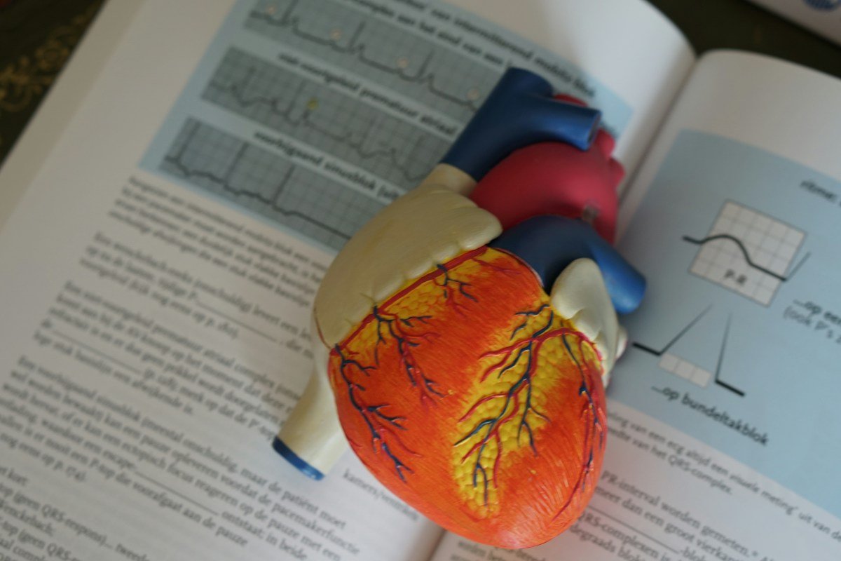

Cardiovascular endothelial function constitutes the foundational physiological determinant of human vascular longevity, arterial elasticity, and systemic organ perfusion. Spanning an estimated surface area equivalent to several tennis courts and lining the entirety of the human circulatory architecture, the vascular endothelium is not an inert biological conduit; rather, it is a metabolically dynamic autocrine, paracrine, and endocrine organ. Weighing over one kilogram in an adult human, this cellular monolayer governs vascular smooth muscle tone, modulates platelet aggregation, regulates leukocyte extravasation, controls trans-endothelial nutrient exchange, and directs local inflammatory responses.
At the molecular vanguard of vascular health operates the endothelial nitric oxide synthase (eNOS) enzymatic pathway. The continuous, pulsatile synthesis of endogenous nitric oxide (NO) from L-arginine confers essential vasoprotective properties: promoting profound vasodilation, suppressing vascular smooth muscle cell proliferation, blunting leukocyte adhesion, and preventing intravascular thrombogenesis. Conversely, endothelial dysfunction—characterized by uncoupled eNOS, diminished nitric oxide bioavailability, accelerated oxidative degradation via superoxide anions, and heightened expression of vascular cell adhesion molecules—serves as the primary initiating event in atherosclerosis, arterial stiffening, hypertension, and microvascular ischemia.
Modern cardiovascular medicine increasingly recognizes that chronological aging does not dictate arterial stiffness; rather, biological vascular age is dictated by the biochemical integrity of the endothelial glycocalyx and the enzymatic fidelity of nitric oxide synthesis. Optimizing endothelial function requires moving beyond conventional pharmacotherapies toward a multi-systemic physiological intervention: exploiting fluid shear stress kinetics through structured resistance and aerobic exercise, entraining tetrahydrobiopterin (BH4) cofactor recycling, preserving endothelial glycocalyx architecture, leveraging dietary nitrate-nitrite-NO enterosalivary cascades, and minimizing vascular advanced glycation end-products.
This comprehensive clinical and biochemical guide delivers an authoritative, evidence-based masterclass in cardiovascular endothelial optimization. Designed for cardiologists, vascular physiologists, and dedicated health optimizers, this manual translates cutting-edge vascular biology into clinically actionable protocols. By systematically mastering the hemodynamic, nutritional, and physical interventions that govern endothelial compliance, practitioners can effectively arrest arterial aging, enhance systemic tissue micro-perfusion, and construct impenetrable cardiovascular resilience.
Endothelial Cell Biology and Glycocalyx Architecture: The Protective Surface Matrix
The vascular endothelium comprises approximately sixty trillion squamoid endothelial cells lining the luminal surface of every artery, vein, capillary, and lymph vessel. The luminal interface of each endothelial cell is shielded by a delicate, carbohydrate-rich polymeric meshwork known as the endothelial glycocalyx. Extending several hundred nanometers into the vascular lumen, this negatively charged surface matrix consists of membrane-bound proteoglycans (syndecans and glypicans) decorated with glycosaminoglycan side chains: heparan sulfate, chondroitin sulfate, and high-molecular-weight hyaluronic acid.
The glycocalyx operates as the vascular system’s primary biophysical barrier and sensory transducer. It repels circulating negatively charged red blood cells, platelets, and leukocytes, preventing mechanical abrasion and pathological cellular adhesion against the fragile endothelial plasma membrane. Furthermore, the glycocalyx incorporates antioxidant enzymes, specifically extracellular superoxide dismutase (ecSOD), which neutralizes flowing reactive oxygen species before they can degrade local nitric oxide.
Crucially, the proteoglycan core proteins of the glycocalyx are mechanically coupled to the intracellular actin cytoskeleton of the endothelial cell. When flowing blood exerts frictional drag across the luminal surface, the glycocalyx bends, transmitting mechanical forces into intracellular focal adhesion complexes. This mechanotransduction cascade is the obligate physical trigger that commands endothelial nitric oxide synthase to synthesize and release nitric oxide into the arterial wall.
Disruption of the endothelial glycocalyx—provoked by postprandial hyperglycemia, hypernatremia, oxidized LDL, endotoxemia, or systemic inflammation—strips this protective layer. Glycocalyx shedding exposes surface adhesion molecules (VCAM-1 and ICAM-1), invites monocyte infiltration into the sub-endothelial intima, and impairs mechanotransduction-mediated vasodilation, marking the initial histological stage of atherogenesis.
Biochemical Kinetics of Endothelial Nitric Oxide Synthase (eNOS) and L-Arginine Metabolism
Endothelial nitric oxide synthase (eNOS) is a complex, multi-domain homodimeric enzyme anchored to caveolae on the inner surface of the endothelial plasma membrane. Each eNOS monomer consists of an amino-terminal oxygenase domain—possessing binding sites for heme, tetrahydrobiopterin (BH4), and the substrate L-arginine—and a carboxy-terminal reductase domain containing binding sites for flavin mononucleotide (FMN), flavin adenine dinucleotide (FAD), and NADPH.
Under physiological conditions, shear stress or hormonal agonists (such as bradykinin or acetylcholine) trigger an influx of intracellular calcium ions. Calcium binds to calmodulin, inducing a conformational change that displaces caveolin-1 and allows calmodulin to bind between the reductase and oxygenase domains. This binding facilitates the transfer of electrons from NADPH through FAD and FMN to the catalytic heme center, where molecular oxygen oxidizes the guanidino nitrogen of L-arginine to synthesize nitric oxide and stoichiometric quantities of L-citrulline.
Synthesized nitric oxide, an uncharged hydrophobic gas with a biological half-life of mere seconds, diffuses rapidly across the internal elastic lamina into adjacent vascular smooth muscle cells. Within smooth muscle cytoplasm, nitric oxide binds to the heme moiety of soluble guanylyl cyclase (sGC), accelerating its enzymatic activity by several hundred fold. Active sGC catalyzes the conversion of guanosine triphosphate (GTP) into cyclic guanosine monophosphate (cGMP).
Accumulating intracellular cGMP activates Protein Kinase G (PKG), which orchestrates vascular smooth muscle relaxation through three distinct mechanisms: phosphorylating phospholamban to stimulate sarcoplasmic reticulum calcium ATPase (SERCA) calcium reuptake, inhibiting voltage-gated L-type calcium channels, and stimulating myosin light chain phosphatase to dephosphorylate myosin light chains. The resulting fall in free cytosolic calcium and cessation of cross-bridge cycling produces robust, rapid vasodilation.
The Tetrahydrobiopterin (BH4) Redox Molecular Switch and eNOS Uncoupling
The enzymatic fidelity of eNOS hinges entirely upon the availability of its essential redox cofactor: (6R)-5,6,7,8-tetrahydrobiopterin (BH4). BH4 acts as an essential molecular glue that stabilizes the functional eNOS homodimer and supplies the single electron required to activate the ferrous-dioxygen complex during substrate oxidation.
Under conditions of sustained oxidative stress, elevated intracellular concentrations of superoxide anions (O2.-) react with native nitric oxide at diffusion-limited rates (approximately 1.9 x 10^10 M^-1 s^-1), generating the highly cytotoxic oxidant peroxynitrite (ONOO-). Peroxynitrite rapidly oxidizes BH4 into biologically inactive dihydrobiopterin (BH2) and biopterin. Deprived of adequate BH4, the eNOS homodimer uncouples into inactive monomers.
When uncoupled eNOS transfers electrons from NADPH, it can no longer successfully reduce L-arginine. Instead, electrons leak directly onto molecular oxygen, synthesizing additional superoxide anions rather than nitric oxide. This catastrophic functional inversion transforms eNOS from a premier cardioprotective vasorelaxant enzyme into an autonomous generator of destructive oxidative stress.
Rescuing uncoupled eNOS requires increasing the intracellular BH4:BH2 ratio. Therapeutic strategies involve upregulating GTP cyclohydrolase I (GTPCH-1)—the rate-limiting enzyme in de novo BH4 biosynthesis—via shear stress and peroxisome proliferator-activated receptor alpha (PPAR-alpha) agonism, paired with high-dose intracellular antioxidants (such as ascorbate and tetrahydrofolate) that chemically reduce trihydrobiopterin radicals back into active BH4.
Asymmetric Dimethylarginine (ADMA) and the Arginase Competing Pathway
Even when eNOS is fully coupled and furnished with BH4, nitric oxide production can be blunted by endogenous competitive substrate antagonists. The primary endogenous antagonist is Asymmetric Dimethylarginine (ADMA), a modified amino acid generated during the natural post-translational methylation of intranuclear histone proteins by protein arginine methyltransferases (PRMTs).
Upon cellular proteolysis, free ADMA is released into the cytoplasm and systemic circulation, where it competes directly with L-arginine for the active binding pocket of eNOS. Because ADMA binds with equal affinity to eNOS but cannot be oxidized to synthesize nitric oxide, elevated plasma ADMA concentrations (exceeding 0.6 to 0.7 micromoles per liter) act as a potent brake on vascular nitric oxide output, directly predicting future myocardial infarction, stroke, and cardiovascular mortality.
ADMA clearance is governed enzymatically by Dimethylarginine Dimethylaminohydrolase (DDAH), which hydrolyzes ADMA into dimethylamine and L-citrulline. However, DDAH contains a critical catalytic cysteine residue that is exquisitely sensitive to oxidative inactivation by hydrogen peroxide and oxLDL. Thus, systemic oxidative stress indirectly elevates ADMA levels by paralyzing DDAH clearance mechanisms.
Simultaneously, the enzyme arginase (both cytosolic arginase-1 and mitochondrial arginase-2) competes with eNOS for their mutual substrate, L-arginine. Arginase metabolizes L-arginine into L-ornithine and urea. In vascular inflammation, arginase expression is massively upregulated by TNF-alpha and oxLDL, starving eNOS of intracellular L-arginine (a phenomenon termed the L-Arginine Paradox) and shunting ornithine into polyamine and proline synthesis, which drives vascular smooth muscle hypertrophy and collagenous arterial stiffening.
Hemodynamic Shear Stress Mechanics: Laminar Versus Oscillatory Fluid Dynamics
Fluid shear stress—the frictional tangential force exerted by flowing blood against the apical surface of endothelial cells—is the principal physiological regulator of vascular phenotype and gene expression. Measured in dynes per square centimeter (dyn/cm2), shear stress characteristics dictate whether the endothelium expresses an atheroprotective or atheroprone gene profile.
High unidirectional laminar shear stress (ranging from 15 to 40 dyn/cm2), encountered in straight arterial segments during physical activity, stimulates endothelial mechanosensory complexes (PECAM-1, VE-cadherin, and VEGFR2). Mechanotransduction activates the Kruppel-Like Factor 2 (KLF2) and Kruppel-Like Factor 4 (KLF4) transcriptional master regulators. KLF2 commands the sustained transcription of eNOS, thrombomodulin, and extracellular superoxide dismutase, while potently suppressing the NF-kB pathway, VCAM-1, and endothelin-1.
In stark contrast, low or oscillatory shear stress (characterized by turbulent, reversing flow patterns averaging less than 4 dyn/cm2) occurs naturally at arterial bifurcations, curvatures, and branching points, as well as in sedentary individuals with low cardiac output. Oscillatory flow fails to stimulate KLF2; instead, it activates the transcription factors AP-1 and NF-kB, driving endothelial cell apoptosis, increasing macromolecular permeability, and transforming the endothelium into a pro-atherogenic, sticky surface.
Exercise prescription for cardiovascular longevity is designed fundamentally to convert systemic vascular beds from low, stagnant shear states into high-velocity pulsatile laminar shear environments. Repeated bouts of laminar shear stress induce structural arterial remodeling (outward arterial lumen enlargement) and enhance basal endothelial nitric oxide synthetic capacity across the entire circulatory tree.
Arterial Elasticity, Pulse Wave Velocity, and Conduit Vessel Compliance
Large central elastic arteries, particularly the thoracic aorta and carotid arteries, function as a biological hydraulic capacitor, termed the Windkessel effect. During ventricular systole, compliant aortic walls distend to absorb approximately fifty percent of the left ventricular stroke volume, converting kinetic hydraulic energy into potential elastic energy. During ventricular diastole, passive elastic recoil of the aortic wall propels this stored blood forward into coronary and peripheral capillary beds, maintaining continuous microvascular perfusion while dampening pulse pressure.
With advancing vascular age and chronic endothelial dysfunction, arterial walls undergo pathological remodeling: resilient, highly elastic elastin fibers are progressively fragmented by matrix metalloproteinases (MMP-2 and MMP-9), replaced by stiff, unyielding type I and type III collagen cross-linked by advanced glycation end-products. Concurrently, loss of basal endothelial nitric oxide increases the passive myogenic tone of vascular smooth muscle cells.
Arterial stiffness is quantified clinically via Carotid-Femoral Pulse Wave Velocity (cfPWV), the recognized gold-standard metric of aortic stiffness. In compliant, elastic youthful vessels, the systolic pressure wave travels at a leisurely speed of five to seven meters per second (m/s). As arteries stiffen, the pulse wave velocity accelerates dramatically, exceeding ten to twelve meters per second.
Elevated cfPWV carries catastrophic hemodynamics consequences: the forward pulse wave travels so rapidly that its reflection from peripheral vascular bifurcations returns to the ascending aorta during late systole rather than diastole. This reflected wave augments peak systolic blood pressure (increasing left ventricular afterload, myocardial oxygen demand, and promoting left ventricular hypertrophy) while precipitating a precipitous drop in diastolic pressure, starving coronary capillary networks that depend strictly upon diastolic perfusion.
Resistance Training and Endothelial Shear Kinetics: Vascular Shear Patterns
The impact of physical exercise on vascular health has historically been dominated by aerobic endurance research. However, modern vascular kinesiology demonstrates that structured progressive resistance training exerts profound, unique hemodynamic shear stress stimuli upon peripheral and central vascular beds.
During the concentric contraction phase of high-intensity resistance training (such as a heavy squat or leg press), intramuscular pressure surges, mechanically compressing muscular arterioles and temporarily interrupting forward arterial inflow. Concurrently, high intra-thoracic pressures generated during the Valsalva maneuver transiently elevate arterial blood pressure.
During the subsequent eccentric phase and immediate post-set relaxation, intramuscular pressure collapses, triggering a massive hemodynamic phenomenon known as reactive hyperemia. Blood rushes into the dilated peripheral vascular bed under immense pressure gradients, generating turbulent, high-velocity forward laminar shear stress across arterial conduits that exceeds resting shear rates by five to ten fold. This intense shear pulse strongly activates endothelial mechanoreceptors, inducing acute phosphorylation of eNOS at Serine 1177 via the Akt/PI3K kinase pathway.
When programmed with moderate intensities (sixty to seventy-five percent of one-repetition maximum), controlled tempos, and adequate rest intervals, resistance training enhances microvascular capillary density, stimulates vascular endothelial growth factor (VEGF), increases flow-mediated dilation (FMD), and preserves skeletal muscle mass—the primary metabolic sink for glucose disposal that prevents postprandial endothelial glycation.
Aerobic Conditioning and High-Intensity Interval Training for Microvascular Density
While resistance training delivers intermittent hyperemic shear spikes, continuous aerobic conditioning and High-Intensity Interval Training (HIIT) deliver sustained, systemic increases in cardiac output that elevate mean arterial shear stress for extended durations.
Zone 2 steady-state aerobic conditioning (sustained continuous exercise performed below the first ventilatory threshold, where blood lactate remains below 2.0 millimoles per liter) provides uninterrupted, high-volume laminar shear stress across central and peripheral conduits without inducing excessive systemic sympatho-excitation or oxidative fatigue. Performing thirty to forty-five minutes of Zone 2 aerobic cycling or rowing stimulates mitochondrial biogenesis in endothelial cells, upregulates eNOS gene expression, and enhances systemic endothelial glycocalyx thickness.
Complementary to Zone 2, High-Intensity Interval Training (HIIT)—alternating brief intervals of near-maximal effort (eighty-five to ninety-five percent of VO2 max) with passive recovery—stimulates significant improvements in brachial artery flow-mediated dilation. The rapid oscillations between maximal metabolic demand and active recovery trigger transient episodic hypoxia followed by hyper-oxygenation, a powerful hormetic stimulus that mobilizes Endothelial Progenitor Cells (CD34+/KDR+ EPCs) from bone marrow niches into circulation to repair damaged endothelial monolayers and promote coronary angiogenesis.
Dietary Nitrate-Nitrite-Nitric Oxide Enterosalivary Physiological Pathway
In addition to the classical oxygen-dependent L-arginine-eNOS enzymatic pathway, human biology possesses an evolutionary backup system for nitric oxide generation: the oxygen-independent Nitrate-Nitrite-Nitric Oxide enterosalivary pathway. Discovered in modern cardiovascular biochemistry, this pathway operates with supreme efficiency in hypoxic, acidic tissues where native eNOS is severely compromised.
Dietary inorganic nitrate (NO3-), abundant in green leafy vegetables (arugula, spinach, Swiss chard) and red beetroot (Beta vulgaris), is rapidly absorbed across the upper gastrointestinal tract into systemic circulation. Approximately twenty-five percent of circulating nitrate is actively extracted by the salivary glands and concentrated up to ten-fold in saliva.
Upon secretion into the oral cavity, facultative anaerobic bacteria residing in deep crypts on the posterior dorsal surface of the tongue (particularly species of Veillonella, Actinomyces, and Rothia) utilize nitrate as an alternative terminal electron acceptor, employing bacterial nitrate reductase enzymes to reduce inorganic nitrate into inorganic nitrite (NO2-). Crucially, the use of commercial antibacterial chlorhexidine mouthwashes destroys this oral microbiome community, abolishing oral nitrite synthesis and precipitating acute systemic hypertension.
Swallowed saliva delivers concentrated nitrite into the acidic gastric juice of the stomach (pH 1.5 to 2.0), where it is instantly protonated into nitrous acid (HNO2), which decomposes into nitric oxide gas and other nitrogen oxides that protect gastric mucosal blood flow. Unreduced nitrite passes into the duodenum, enters systemic circulation, and travels to peripheral vascular beds. In areas of low oxygen tension or acidosis, circulating nitrite is reduced into bioactive nitric oxide by deoxygenated hemoglobin, myoglobin, and endothelial xanthine oxidoreductase, producing targeted vasodilation precisely where tissues require perfusion.
Mitochondrial Dynamics in Endothelial Bioenergetics: Mitophagy and ROS Signaling
Although vascular endothelial cells derive approximately eighty-five percent of their cellular adenosine triphosphate (ATP) from anaerobic glycolysis rather than oxidative phosphorylation, endothelial mitochondria serve as indispensable sensory hubs for intracellular redox homeostasis, calcium buffering, and apoptosis regulation.
Under physiological conditions, healthy endothelial mitochondria undergo continuous, coordinated cycles of fusion (orchestrated by Mitofusin 1, Mitofusin 2, and OPA1) and fission (governed by Dynamin-Related Protein 1, Drp1). Fusion permits the exchange of material between functional mitochondria, while fission isolates depolarized, dysfunctional segments of the mitochondrial reticular network for targeted autophagic degradation, a selective cellular cleanup pathway designated as mitophagy (mediated by the PINK1/Parkin enzymatic axis).
In chronic endothelial dysfunction—accelerated by persistent hyperglycemia, elevated free fatty acids, and inflammatory cytokines—mitochondrial fission surges out of control. Hyperactive Drp1 causes extensive mitochondrial fragmentation, accompanied by excessive generation of mitochondrial reactive oxygen species (mtROS) from respiratory complexes I and III. mtROS oxidizes adjacent cell structures, induces the opening of the mitochondrial permeability transition pore (mPTP), and triggers the cytoplasmic leakage of oxidized mitochondrial DNA, which activates the sterile cGAS-STING inflammasome and commands endothelial apoptosis.
Endothelial-to-Mesenchymal Transition (EndMT) in Vascular Remodeling and Fibrosis
A profoundly disruptive cellular phenomenon driving accelerated vascular stiffening and plaque vulnerability is Endothelial-to-Mesenchymal Transition (EndMT). During EndMT, differentiated, polarized endothelial cells lose their specific endothelial molecular markers, including vascular endothelial cadherin (VE-cadherin) and CD31 (PECAM-1), while abandoning their characteristic cobble-stone morphology.
Under the command of transforming growth factor-beta (TGF-beta), bone morphogenetic protein (BMP) signaling, and inflammatory cytokines (IL-1 beta and TNF-alpha), transitional endothelial cells upregulate mesenchymal transcription factors: Snail, Slug, Twist, and ZEB1. The transformed cells acquire a migratory, spindle-shaped mesenchymal phenotype, expressing high concentrations of alpha-smooth muscle actin (alpha-SMA), vimentin, and type I collagen.
These transformed mesenchymal-like cells delaminate from the endothelial monolayer and migrate into the sub-endothelial intima. Within the intimal space, they actively synthesize dense extracellular matrix proteins, driving progressive luminal narrowing, diffuse fibrotic wall thickening, and calcification. Furthermore, EndMT destabilizes existing atherosclerotic plaques: by stripping the protective fibrous cap of endothelial coverage and producing excess matrix metalloproteinases, EndMT transforms stable plaques into rupture-prone, lethal lesions.
Circadian Rhythms of Endothelial Function and Morning Cardiovascular Vulnerability
Human cardiovascular physiology is governed by strict, evolutionary conserved diurnal circadian rhythms entrained by the suprachiasmatic nucleus (SCN) of the anterior hypothalamus and peripheral molecular clock genes (CLOCK, BMAL1, PER, and CRY) expressed directly within endothelial and vascular smooth muscle cells.
Clinical epidemiological data demonstrates an unmistakable, dangerous peak in acute cardiovascular events—including ST-segment elevation myocardial infarction, sudden cardiac death, and ischemic cerebrovascular stroke—between the morning hours of six o’clock and eleven o’clock AM. This vulnerability window coincides precisely with a physiological nadir in basal endothelial function and nitric oxide bioavailability.
During the early morning awakening period, the cortisol awakening response (CAR) triggers a surge in central sympathetic nervous system outflow, elevating systemic catecholamines (epinephrine and norepinephrine). Concurrently, circulating levels of Plasminogen Activator Inhibitor-1 (PAI-1)—the primary inhibitor of endogenous fibrinolysis—reach their 24-hour circadian zenith, while vascular alpha-1 adrenergic sensitivity increases. Endothelial cells exhibit blunted flow-mediated dilation and impaired shear-stress responsiveness during morning hours, highlighting the imperative of chronotherapeutic scheduling for blood pressure medications, targeted nitrate supplementation, and morning hydration.
Gasotransmitter Cross-Talk: Hydrogen Sulfide and Carbon Monoxide Synergies
While nitric oxide has historically received the lion’s share of cardiovascular research, contemporary vascular biology recognizes that endothelial tone is governed by an interdependent triumvirate of endogenous gaseous signaling molecules, collectively termed gasotransmitters: Nitric Oxide (NO), Hydrogen Sulfide (H2S), and Carbon Monoxide (CO).
Hydrogen sulfide is synthesized within vascular endothelial and smooth muscle cells primarily by cystathionine gamma-lyase (CSE) and 3-mercaptopyruvate sulfurtransferase (3-MST) utilizing L-cysteine and homocysteine as substrates. Unlike nitric oxide, which signals primarily via soluble guanylyl cyclase and cGMP, hydrogen sulfide directly activates ATP-sensitive potassium channels (K_ATP channels) on vascular smooth muscle membranes. The resulting potassium efflux hyperpolarizes the smooth muscle cell membrane, closing voltage-gated calcium channels and causing profound Endothelium-Dependent Hyperpolarization (EDH)-mediated vasodilation.
Crucially, hydrogen sulfide and nitric oxide exhibit intense biochemical synergy. Hydrogen sulfide sulfhydrates (S-sulfenylates) critical cysteine residues on the eNOS enzyme, stabilizing its active homodimeric conformation and preventing uncoupling. Simultaneously, physiological concentrations of H2S inhibit phosphodiesterase-5 (PDE-5), preventing the enzymatic degradation of cGMP and amplifying nitric oxide-mediated vasorelaxation. When endogenous H2S production declines—whether due to micronutrient deficiencies in vitamin B6 (an obligate cofactor for CSE) or oxidative stress—nitric oxide responsiveness collapses.
Carbon monoxide, synthesized by endothelial heme oxygenase-1 (HO-1) during the catabolism of heme into biliverdin and iron, acts as an additional endogenous brake on vascular inflammation. HO-1-derived CO binds to soluble guanylyl cyclase to stimulate cGMP synthesis, while exerting potent anti-apoptotic and anti-thrombotic protections across the endothelial monolayer. Supporting endogenous gasotransmitter synthesis through sulfur-rich allium vegetables (garlic, shallots), N-acetylcysteine, and Nrf2-mediated HO-1 induction (via sulforaphane) creates a redundant, resilient trifecta of vasoprotective signaling.
Endothelial Micronutrition: L-Citrulline, Polyphenols, and Methylation Support
Targeted nutritional biochemistry provides clinicians with specialized molecular substrates to enhance endothelial nitric oxide output, scavenge vascular free radicals, and preserve vascular compliance.
While oral L-arginine supplementation has long been marketed for vascular health, oral L-arginine suffers from extensive first-pass hepatic metabolism: up to sixty percent of ingested arginine is extracted and destroyed by hepatic arginases before reaching systemic circulation. In contrast, L-citrulline, a non-protein amino acid abundant in watermelon rind, bypasses hepatic first-pass extraction entirely. Absorbed intact in the intestines, circulating L-citrulline is extracted by renal proximal tubule cells and vascular endothelial cells, which enzymatically convert it into L-arginine via argininosuccinate synthase and argininosuccinate lyase, resulting in far higher and more sustained plasma L-arginine concentrations than equimolar doses of L-arginine itself.
Bioactive polyphenolic compounds deliver critical antioxidant and enzymatic support to vascular endothelium. Oligomeric proanthocyanidins (OPCs) extracted from French maritime pine bark (Pycnogenol) and grape seed extract directly stimulate eNOS phosphorylation while protecting the endothelial glycocalyx from enzymatic degradation by hyaluronidases. Flavan-3-ols abundant in raw non-alkalized cocoa stimulate flow-mediated dilation in humans within two hours of ingestion by blunting NADPH oxidase activity and reducing circulating superoxide.
Furthermore, vascular one-carbon methylation pathways must be meticulously supported. Elevated plasma homocysteine levels (above 10 to 12 micromoles per liter) induce severe endothelial cell apoptosis, oxidatively inactivate DDAH, and decouple eNOS. Clinical optimization demands targeted supplementation with bioavailable methylation cofactors: 5-methyltetrahydrofolate (5-MTHF), methylcobalamin (B12), pyridoxal-5-phosphate (B6), and trimethylglycine (betaine) to convert homocysteine safely into methionine or cysteine.
Intracellular magnesium kinetics play an equally decisive role in maintaining vascular compliance. Acting as nature’s physiological calcium antagonist, elemental magnesium competes directly with ionic calcium for binding sites on troponin C and smooth muscle calmodulin. Intracellular magnesium depletion unleashes unconstrained calcium influx through L-type calcium channels, driving chronic arterial spasm, hypertension, and endothelial oxidative stress. Supplemental organic magnesium chelates—specifically magnesium taurate, which pairs magnesium with the cardioprotective sulfonic acid taurine—suppress endothelin-1 secretion, facilitate eNOS dimerization, and reduce carotid-femoral pulse wave velocity across clinical populations.
Epigenetic post-transcriptional regulation via vascular microRNAs (miRNAs) further controls endothelial longevity. Laminar shear stress stimulates the endothelial expression of microRNA-126 (miR-126) and microRNA-143/145, which act as master atheroprotective regulators by silencing vascular cell adhesion molecule-1 and repressing vascular smooth muscle phenotypic switching. Conversely, sedentary oscillatory shear downregulates miR-126 while elevating pro-inflammatory microRNA-92a, demonstrating that hemodynamic movement patterns directly remodel the molecular genetic operating system of the human arterial wall.
Endothelin-1 Antagonism and Sympathovagal Autonomic Balance
Vascular tone is a continuous balancing act between vasodilating mediators (primarily nitric oxide and prostacyclin) and vasoconstricting mediators, dominant among which is Endothelin-1 (ET-1). Synthesized by endothelial cells as a 21-amino acid peptide, ET-1 is the most potent endogenous vasoconstrictor known in human physiology, exerting vascular contractile effects ten times greater than angiotensin II.
ET-1 acts upon two distinct receptor subtypes: ETA receptors located on vascular smooth muscle cells (which mediate intense, prolonged vasoconstriction and smooth muscle proliferation) and ETB receptors located on endothelial cells (which clear circulating ET-1 and stimulate nitric oxide release). In states of chronic psychological stress, systemic inflammation, or sleep apnea, endothelial ET-1 synthesis surges while endothelial ETB clearance receptors are downregulated, locking systemic vascular beds into chronic hypertension.
Chronically elevated sympathetic nervous system tone accelerates this vasoconstrictive cascade. Continuous adrenergic signaling stimulates alpha-1 adrenergic receptors on peripheral arterioles, generating vascular constriction and high vascular resistance. Interventions that shift the autonomic nervous system toward parasympathetic vagal dominance—including resonance-frequency slow breathing (5.5 to 6 breaths per minute), cold-water facial immersion, and deep slow-wave sleep—suppress sympathetic outflow, blunt circulating catecholamines, and restore the natural balance between nitric oxide and Endothelin-1.
Clinical Diagnostic Methodology: Flow-Mediated Dilation and Tonometry
Objective clinical quantification of endothelial function requires validated, non-invasive biophysical assessment modalities that measure dynamic vascular responsiveness rather than static anatomic plaque.
Flow-Mediated Dilation (FMD) of the brachial artery represents the accepted clinical research gold standard for measuring in vivo endothelial nitric oxide bioavailability. Utilizing high-resolution vascular ultrasound, the clinician measures the baseline diameter of the brachial artery. A pneumatic occlusion cuff placed around the forearm is then inflated to suprasystolic pressure (typically 200 to 250 mmHg) for exactly five minutes, inducing complete distal ischemia.
Upon cuff deflation, the sudden surge in blood flow creates immense laminar shear stress across the brachial artery endothelium. In a healthy vessel, mechanosensitive eNOS activation releases a burst of nitric oxide, causing the artery to dilate by seven to twelve percent within sixty to ninety seconds. An FMD response below five to six percent identifies significant endothelial dysfunction and independently predicts future adverse cardiovascular events.
Complementing FMD, Peripheral Arterial Tonometry (EndoPAT) measures digital pulse wave amplitude before and after reactive hyperemia, generating a Reactive Hyperemia Index (RHI) that reflects microvascular endothelial compliance. SphygmoCor arterial tonometry quantifies Central Aortic Systolic Pressure (CASP), the Augmentation Index (AIx), and Carotid-Femoral Pulse Wave Velocity, providing comprehensive mapping of systemic arterial stiffness.
To translate these advanced vascular principles into clinical protocols, cardiovascular specialists rely on structured diagnostic and intervention matrices. These frameworks systematically map endothelial pathology to molecular mechanisms, objective diagnostic criteria, progressive lifestyle and nutritional interventions, and measurable clinical endpoints.
The following comprehensive comparative clinical matrix provides an authoritative reference evaluating primary endothelial dysfunction phenotypes, their underlying biomolecular drivers, diagnostic metrics, and targeted therapeutic strategies.
Comparative Clinical Matrix of Endothelial Dysfunction Phenotypes & Interventions
| Endothelial Dysfunction Phenotype | Primary Biomolecular Drivers | Clinical Diagnostic Metrics | Targeted Nutritional & Exercise Intervention | Measurable Resolution Endpoints |
|---|---|---|---|---|
| Uncoupled eNOS & Oxidative Stress | BH4 oxidation by peroxynitrite; eNOS monomerization; superoxide leakage | FMD < 5.0%; elevated serum nitrotyrosine; elevated hs-CRP > 2.0 mg/L | L-citrulline (6-8g daily); Pycnogenol (150mg); Zone 2 aerobic training (150 min/wk) | FMD restored > 8.0%; normalization of serum nitrotyrosine; reduced resting pulse |
| Aortic Elasticity Loss / Stiffening | Elastin fragmentation; advanced glycation cross-links; collagen accumulation | cfPWV > 10.0 m/s; central pulse pressure > 50 mmHg; Augmentation Index > 30% | Dietary nitrates (beetroot 400mg NO3-); high-dose cocoa flavanols (900mg); moderate resistance work | cfPWV reduction > 1.5 m/s; central pulse pressure < 40 mmHg; improved coronary reserve |
| Substrate Competition / Hyper-ADMA | DDAH oxidative inhibition; elevated PRMT activity; arginase upregulation | Plasma ADMA > 0.65 umol/L; low L-arginine/ADMA ratio (< 100); elevated homocysteine | Active methylfolate (5-MTHF 1,000mcg) + methyl-B12; N-acetylcysteine (1,200mg); HIIT cycling | ADMA normalized < 0.45 umol/L; homocysteine < 9 umol/L; restoration of exercise tolerance |
| Glycocalyx Degradation / Permeability | Hyperglycemic stripping; matrix metalloproteinase shedding; loss of syndecans | Elevated plasma syndecan-1 (> 50 ng/mL); elevated circulating hyaluronan; microalbuminuria | High-molecular-weight hyaluronic acid; sulodexide; low-glycemic Mediterranean nutrition | Syndecan-1 normalized; elimination of microalbuminuria; restored shear-mediated mechanosensation |
| Sympathetic Hypertonicity / Vasospasm | Endothelin-1 excess; alpha-1 adrenergic vasoconstriction; vagal withdrawal | EndoPAT RHI < 1.67; low HRV (RMSSD < 25 ms); elevated nocturnal blood pressure dipping deficit | Resonance-frequency breathing (0.1 Hz); magnesium glycinate (400mg); Finnish sauna (80C, 20 min) | EndoPAT RHI > 2.10; RMSSD doubled; restoration of physiological nocturnal dipping (> 10%) |
Implementing these comprehensive vascular interventions enables healthcare professionals to systematically reverse endothelial senescence and optimize arterial compliance. For authoritative research on vascular biology, nitric oxide pharmacology, and clinical cardiology, practitioners consult established global institutions including the National Heart, Lung, and Blood Institute Vascular Research Division and the Circulation Academic Journal Collection. Additional microvascular research can be reviewed through the Cardiovascular Research European Journal Portal, alongside hypertension guidelines curated by the European Society of Cardiology Guidelines Portal and endothelial diagnostics standards from the American College of Cardiology Clinical Library.
Frequently Asked Questions About Cardiovascular Endothelial Function
What is the primary role of the vascular endothelium?
The vascular endothelium is an active endocrine and paracrine organ that lines the entire circulatory system. It regulates vascular tone through nitric oxide synthesis, prevents pathological blood clotting, modulates inflammation, controls vascular permeability, and prevents leukocyte adhesion to arterial walls.
What causes endothelial nitric oxide synthase (eNOS) uncoupling?
eNOS uncoupling is triggered primarily by the oxidative degradation of its essential cofactor, tetrahydrobiopterin (BH4), into inactive dihydrobiopterin (BH2) by peroxynitrite. Without BH4, the eNOS homodimer uncouples, producing destructive superoxide radicals instead of protective nitric oxide.
Why is L-citrulline superior to L-arginine for boosting nitric oxide?
Oral L-arginine suffers extensive first-pass breakdown by arginase enzymes in the liver and intestines, with up to sixty percent eliminated before reaching systemic blood. L-citrulline completely bypasses hepatic first-pass metabolism and is converted into L-arginine directly within the kidneys and vascular endothelium, yielding higher and more sustained plasma arginine levels.
How does physical exercise improve arterial compliance?
Exercise increases cardiac output, generating high pulsatile laminar shear stress across arterial walls. This mechanical drag stimulates mechanoreceptors, activating KLF2 transcription factors and phosphorylating eNOS. Over time, exercise stimulates vascular remodeling, enhances capillary density, and preserves elastin architecture.
What is Flow-Mediated Dilation (FMD) and how is it measured?
Flow-Mediated Dilation is the clinical gold standard for measuring endothelial function. Using vascular ultrasound, clinicians measure the diameter of the brachial artery before and after five minutes of forearm occlusion. Healthy endothelium responds to the rush of blood by dilating seven to twelve percent via nitric oxide release; a response below five percent indicates endothelial dysfunction.
How do antibacterial mouthwashes cause elevated blood pressure?
Antibacterial mouthwashes destroy facultative anaerobic bacteria residing on the dorsal surface of the tongue. These bacteria are responsible for reducing dietary inorganic nitrate into nitrite, the critical step in the enterosalivary nitric oxide pathway. Eliminating these oral microbes blunts systemic nitrite levels, increasing systemic vascular resistance and raising blood pressure.
What is Pulse Wave Velocity (PWV) and what does it indicate?
Carotid-Femoral Pulse Wave Velocity measures the speed at which arterial pressure waves travel through the aorta. In youthful, elastic arteries, waves travel slowly (five to seven meters per second). In stiffened, diseased arteries, pulse wave velocity exceeds ten meters per second, indicating loss of arterial compliance and predicting elevated cardiovascular risk.
How does Endothelin-1 counteract nitric oxide?
Endothelin-1 (ET-1) is the most potent vasoconstricting peptide produced by endothelial cells, exerting ten times the vasoconstrictive power of angiotensin II. In healthy states, nitric oxide suppresses ET-1 synthesis. When the endothelium is damaged, ET-1 synthesis surges, binding to ETA receptors on smooth muscle and causing prolonged, pathological arterial constriction.
What is the endothelial glycocalyx and why is it vital?
The endothelial glycocalyx is a carbohydrate-rich, gel-like meshwork of proteoglycans and glycosaminoglycans coating the luminal surface of endothelial cells. It repels blood cells and platelets to prevent clotting, harbors antioxidant enzymes, and acts as the primary mechanical sensor transmitting fluid shear stress into nitric oxide production.
Cardiovascular Endothelial Synthesis and Lifelong Arterial Resilience
Cardiovascular health and biological longevity are fundamentally bounded by the functional integrity of the vascular endothelium. By understanding the intricate molecular balance between eNOS coupling, tetrahydrobiopterin preservation, laminar shear stress mechanotransduction, and dietary nitrate pathways, clinicians and individuals can systematically eliminate arterial stiffening and microvascular decline. When reinforced through targeted resistance training, Zone 2 aerobic conditioning, L-citrulline supplementation, and autonomic vagal entrainment, the human vascular network retains youthful elasticity, ensuring decades of vibrant tissue perfusion, cardiovascular vitality, and complete protection against degenerative vascular pathology.